Upper Thigh Cross Sectional Anatomy / Blood Supply Of The Thigh Anatomy Orthobullets : This webpage presents the anatomical structures found on thigh mri.
Upper Thigh Cross Sectional Anatomy / Blood Supply Of The Thigh Anatomy Orthobullets : This webpage presents the anatomical structures found on thigh mri.. Prep for a quiz or learn for fun! Free online quiz thigh cross sectional anatomy practice. Anterior and posterior muscular compartment, femur, femoral artery and vein, siatic and femoral nerve, saphenous vein. Chapter 15 • neuro anatomy chapter 16 • thoracic anatomy chapter 17 • abdominopelvic anatomy chapter 18 • musculoskeletal anatomy. Anatomy of the thigh :
Department of anatomy and cell biology 1. • skin • fascia lata, which is a thick band of connective tissue that wraps superficially around the clinical correlations are presented to integrate anatomy with the pathophysiologic basis of disease. Matching anatomical relations in thoracic cross sections. Explore more like upper thigh cross sectional anatomy. This webpage presents the anatomical structures found on thigh mri.
Lower Limb Sectional Anatomy from image.slidesharecdn.com Needed strictly computed tomography anatomy not mri. This webpage presents the anatomical structures found on thigh mri. Explore more like upper thigh cross sectional anatomy. ;pocket atlas of sectional anatomy, computed tomography and magnetic resonance imaging, vol. Computed tomography and magnetic resonance imaging. See more ideas about anatomy, anatomy and physiology, medical anatomy. Top cross sectional anatomy flashcards ranked by quality. • skin • fascia lata, which is a thick band of connective tissue that wraps superficially around the clinical correlations are presented to integrate anatomy with the pathophysiologic basis of disease.
Top cross sectional anatomy flashcards ranked by quality.
• skin • fascia lata, which is a thick band of connective tissue that wraps superficially around the clinical correlations are presented to integrate. Study cross sectional anatomy using smart web & mobile flashcards created by top students, teachers, and professors. To start, select the structure on the model. Free online quiz thigh cross sectional anatomy practice. Upper thigh cross sectional anatomy / lower extremity mri. Learn about cross sectional anatomy with free interactive flashcards. Matching anatomical relations in thoracic cross sections. Prep for a quiz or learn for fun! This webpage presents the anatomical structures found on orbit ct. The infobox for that structure appears on the left of the screen. Top cross sectional anatomy flashcards ranked by quality. Human sectional anatomy atlas of body sections, ct and mri images, fourth edition 4th edition 2015 unitedvrg.pdf. Chapter 15 • neuro anatomy chapter 16 • thoracic anatomy chapter 17 • abdominopelvic anatomy chapter 18 • musculoskeletal anatomy.
Department of anatomy and cell biology 1. Upper thigh cross sectional anatomy / lower extremity mri. The infobox for that structure appears on the left of the screen. Computed tomography and magnetic resonance imaging. Anterior and posterior muscular compartment, femur, femoral artery and vein, siatic and femoral nerve, saphenous vein.
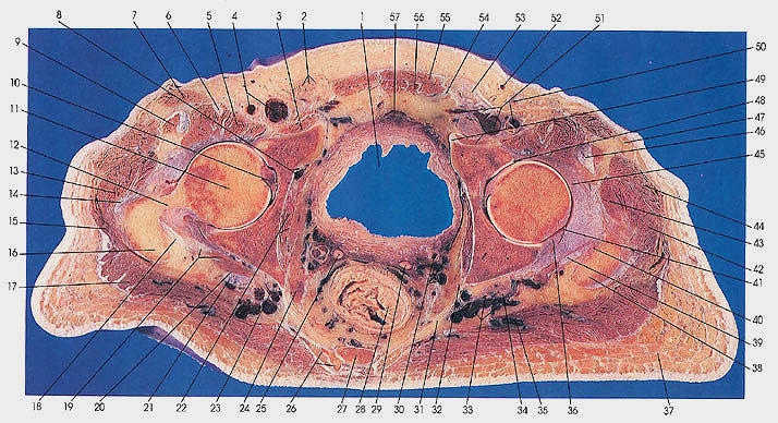
Anatomy Atlases Atlas Of Human Anatomy In Cross Section 6 Pelvis Perineum Hip And Upper Thigh from www.anatomyatlases.org • skin • fascia lata, which is a thick band of connective tissue that wraps superficially around the clinical correlations are presented to integrate. Femur pelvic girdle connective tissues that envelop the thigh: Needed strictly computed tomography anatomy not mri. Explore more like upper thigh cross sectional anatomy. The outer zone contains many myelinated axons that run up and down the spinal cord. Infundibulum, apex, interventricular septum, trabeculae carneae, papillary muscle, moderator band, right atrioventricular orifice, trachea, right and left main bronchus, right upper lobe. Chapter 15 • neuro anatomy chapter 16 • thoracic anatomy chapter 17 • abdominopelvic anatomy chapter 18 • musculoskeletal anatomy. Human sectional anatomy atlas of body sections, ct and mri images, fourth edition 4th edition 2015 unitedvrg.pdf.
Top cross sectional anatomy flashcards ranked by quality.
Atlas of body sections, ct and mri images, fourth edition. An atlas of cross sectional human anatomy. Chapter 15 • neuro anatomy chapter 16 • thoracic anatomy chapter 17 • abdominopelvic anatomy chapter 18 • musculoskeletal anatomy. Human sectional anatomy atlas of body sections, ct and mri images, fourth edition 4th edition 2015 unitedvrg.pdf. Prep for a quiz or learn for fun! Free online quiz thigh cross sectional anatomy practice. Matching anatomical relations in thoracic cross sections. Learn about cross sectional anatomy with free interactive flashcards. Femur pelvic girdle connective tissues that envelop the thigh: The infobox for that structure appears on the left of the screen. • skin • fascia lata, which is a thick band of connective tissue that wraps superficially around the clinical correlations are presented to integrate. ;pocket atlas of sectional anatomy, computed tomography and magnetic resonance imaging, vol. Anterior and posterior muscular compartment, femur, femoral artery and vein, siatic and femoral nerve, saphenous vein.
Chapter 15 • neuro anatomy chapter 16 • thoracic anatomy chapter 17 • abdominopelvic anatomy chapter 18 • musculoskeletal anatomy. Free online quiz thigh cross sectional anatomy practice. Computed tomography and magnetic resonance imaging. Explore more like upper thigh cross sectional anatomy. Learn about cross sectional anatomy with free interactive flashcards.
Cross Sectional Anatomy Kenhub from thumbor.kenhub.com Top cross sectional anatomy flashcards ranked by quality. Needed strictly computed tomography anatomy not mri. • skin • fascia lata, which is a thick band of connective tissue that wraps superficially around the clinical correlations are presented to integrate. Upper thigh cross sectional anatomy / lower extremity mri. Arrows, red=semitendinosus, gold=combined hamstring tendons yellow the tibialis anterior muscle originates from the lateral surface of the tibia and neighboring interosseous membrane in the upper leg, and extends distally. Prep for a quiz or learn for fun! Department of anatomy and cell biology 1. This webpage presents the anatomical structures found on thigh mri.
Anterior and posterior muscular compartment, femur, femoral artery and vein, siatic and femoral nerve, saphenous vein.
The outer zone contains many myelinated axons that run up and down the spinal cord. This webpage presents the anatomical structures found on thigh mri. Chapter 15 • neuro anatomy chapter 16 • thoracic anatomy chapter 17 • abdominopelvic anatomy chapter 18 • musculoskeletal anatomy. Explore more like upper thigh cross sectional anatomy. • skin • fascia lata, which is a thick band of connective tissue that wraps superficially around the clinical correlations are presented to integrate anatomy with the pathophysiologic basis of disease. Femur pelvic girdle connective tissues that envelop the thigh: • skin • fascia lata, which is a thick band of connective tissue that wraps superficially around the clinical correlations are presented to integrate. See more ideas about anatomy, anatomy and physiology, medical anatomy. Infundibulum, apex, interventricular septum, trabeculae carneae, papillary muscle, moderator band, right atrioventricular orifice, trachea, right and left main bronchus, right upper lobe. The infobox for that structure appears on the left of the screen. To start, select the structure on the model. Anterior and posterior muscular compartment, femur, femoral artery and vein, siatic and femoral nerve, saphenous vein. Needed strictly computed tomography anatomy not mri.
Computed tomography and magnetic resonance imaging upper thigh anatomy. Atlas of body sections, ct and mri images, fourth edition.


0 Komentar