Upper Leg Tendon Anatomy - Anterior Thigh : The posterior talofibular ligament is attached to the posterolateral tubercle, which is larger and more prominent than the posteromedial tubercle.
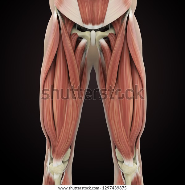
Upper Leg Tendon Anatomy - Anterior Thigh : The posterior talofibular ligament is attached to the posterolateral tubercle, which is larger and more prominent than the posteromedial tubercle.. Originates from the lateral condyle of the tibia and the medial surface of the fibula. Tendons are fibrous cords attached to muscles and bone. Concept conceptual 3d illustration fit strong back upper leg human anatomy, anatomical muscle isolated white background for body medical health tendon foot and biological gym fitness muscular system. Palmar region , arteries (illustrations: Superficial veins of upper limb , anatomy :
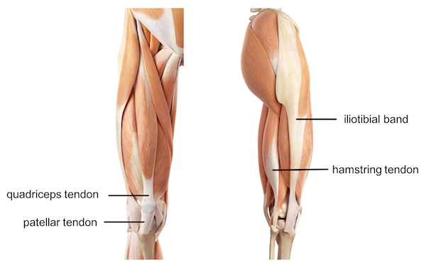
There are four muscles in the anterior compartment of the leg. Originates from the lateral condyle of the tibia and the medial surface of the fibula. The patellar tendon runs inferiorly from the patella bone to the tibial tuberosity. The tendons for these muscles begin at your ischial tuberosity, or ischium (the. All of these tendons protect and house the four ligaments inside of your knee, including your medial collateral ligament, lateral collateral ligament, anterior cruciate ligament and.
Upper Legs Muscles Anatomy 3d Rendering Stock Illustration 1297439875 from image.shutterstock.com Tendons are fibrous cords attached to muscles and bone. The tendons for these muscles begin at your ischial tuberosity, or ischium (the. Use the mouse scroll wheel to move the images up and down alternatively use the tiny arrows (>>) on both side of the image to move the images. Lateral (fibular) collateral ligament (fcl) upper part middle part lower part popliteus tendon (pt) upper part i. Note that the left gastric artery also gives off an oesophageal branch, which passes through the oesophageal hiatus of the diaphragm to supply the lower oesophagus. Palmar region , arteries (illustrations: The sulcus for this tendon is flanked by the posterolateral and posteromedial tubercles. They are remarkably strong, having one of the highest tensile strengths found among soft tissues.
Upper leg anatomy and function.
Hands are outstretched, holding onto the handles of the bench. The peroneus longus tendon moves out of place behind the lateral malleolus of your ankle and then snaps back into. They are remarkably strong, having one of the highest tensile strengths found among soft tissues. Collectively, they act to dorsiflex and invert the foot at the ankle joint. The pads of the machine are situated at the achilles tendon. The achilles tendon or heel cord, also known as the calcaneal tendon, is a tendon at the back of the lower leg, and is the thickest in the human body. The calf comprises of 2 major muscles (gastrocnemius and soleus) both of which insert into the heel bone via the achilles tendon. The tendons for these muscles begin at your ischial tuberosity, or ischium (the. Tendons are fibrous cords attached to muscles and bone. Spicermanyt at checkout for 40% off this tutorial! Fascia of the upper limb. Lie prone on a hamstring curl machine. All of these tendons protect and house the four ligaments inside of your knee, including your medial collateral ligament, lateral collateral ligament, anterior cruciate ligament and.
Hands are outstretched, holding onto the handles of the bench. It serves to attach the plantaris, gastrocnemius (calf) and soleus muscles to the calcaneus (heel) bone. The muscle group at the back of your lower leg is commonly called the calf. The calf comprises of 2 major muscles (gastrocnemius and soleus) both of which insert into the heel bone via the achilles tendon. Spicermanyt at checkout for 40% off this tutorial!
Leg Knee Anatomy from uploads-ssl.webflow.com Lateral (fibular) collateral ligament (fcl) upper part middle part lower part popliteus tendon (pt) upper part i. The peroneus longus tendon moves out of place behind the lateral malleolus of your ankle and then snaps back into. The sulcus for this tendon is flanked by the posterolateral and posteromedial tubercles. Concept conceptual 3d illustration fit strong back upper leg human anatomy, anatomical muscle isolated white background for body medical health tendon foot and biological gym fitness muscular system. Suspensory ligament of the axilla. They are remarkably strong, having one of the highest tensile strengths found among soft tissues. Upper limb trauma programme injuries. Figure 2 posterior relations of the stomach.
This mri wrist coronal cross sectional anatomy tool is absolutely free to use.
Muscle/tendon inflammation and pain along anterio… Suspensory ligament of the axilla. Related online courses on physioplus. Lateral (fibular) collateral ligament (fcl) upper part middle part lower part popliteus tendon (pt) upper part i. Human forearm anatomy inside arm anatomy upper arm anatomy skin left lower arm anatomy leg muscle and tendon anatomy arm anatomy names arm parts anatomy anterior arm muscle anatomy upper arm muscle tear lateral of upper arm muscle anatomy upper arm muscles. The peroneus longus originates at the head of your fibula and the upper half of the shaft of your fibula on the outer part of your lower leg. 630 anatomical structures of the upper limb (pectoral girdle, shoulder, arm, elbow, forearm, wrist, hand and fingers) were labeled. This mri wrist coronal cross sectional anatomy tool is absolutely free to use. (from gray's anatomy 40th edition). Tendon, tissue that attaches a muscle to other body parts, usually bones. .16 penile numbness and perineum tenderness.18 any suggested exercises or stretches?.22 leg musculature 209 elbow tendonitis and saddle sores. Collectively, they act to dorsiflex and invert the foot at the ankle joint. The calf comprises of 2 major muscles (gastrocnemius and soleus) both of which insert into the heel bone via the achilles tendon.
Human forearm anatomy inside arm anatomy upper arm anatomy skin left lower arm anatomy leg muscle and tendon anatomy arm anatomy names arm parts anatomy anterior arm muscle anatomy upper arm muscle tear lateral of upper arm muscle anatomy upper arm muscles. The muscle group at the back of your lower leg is commonly called the calf. • flatfoot deformity • flexible hindfoot • normal forefoot. Collectively, they act to dorsiflex and invert the foot at the ankle joint. The achilles tendon or heel cord, also known as the calcaneal tendon, is a tendon at the back of the lower leg, and is the thickest in the human body.
Drawings Upper Legs Muscles Anatomy Stock Illustration Gg73925215 Gograph from comps.gograph.com This may result in tendon subluxation; Figure 2 posterior relations of the stomach. Note that the left gastric artery also gives off an oesophageal branch, which passes through the oesophageal hiatus of the diaphragm to supply the lower oesophagus. Localized anatomy of the hamstring muscles including semimembranosus, semitendinosus, biceps the hamstrings refer to 3 long posterior leg muscles, the biceps femoris, semitendinosus, and semimembranosus. It serves to attach the plantaris, gastrocnemius (calf) and soleus muscles to the calcaneus (heel) bone. Collectively, they act to dorsiflex and invert the foot at the ankle joint. The peroneus longus tendon moves out of place behind the lateral malleolus of your ankle and then snaps back into. An anatomical and biomechanical study.
Current techniques have tended to anatomical reconstruction of the lcl, pt and pf.
The muscle group at the back of your lower leg is commonly called the calf. Bronchopulmonary segmental anatomy describes the division of the lungs into segments based on the tertiary or segmental bronchi. Palmar region , arteries (illustrations: Fascia of the upper limb. Upper leg anatomy and function. Muscle/tendon inflammation and pain along anterio… 630 anatomical structures of the upper limb (pectoral girdle, shoulder, arm, elbow, forearm, wrist, hand and fingers) were labeled. It serves to attach the plantaris, gastrocnemius (calf) and soleus muscles to the calcaneus (heel) bone. .16 penile numbness and perineum tenderness.18 any suggested exercises or stretches?.22 leg musculature 209 elbow tendonitis and saddle sores. Note that the left gastric artery also gives off an oesophageal branch, which passes through the oesophageal hiatus of the diaphragm to supply the lower oesophagus. Collectively, they act to dorsiflex and invert the foot at the ankle joint. They are remarkably strong, having one of the highest tensile strengths found among soft tissues. A tendon is the fibrous tissue that attaches muscle to bone in the human body.


0 Komentar